Contact Us
Research
-
Seeing the Brain Through the Eye: Multimodal Diagnostic Eye Biomarkers of Cognitive Impairment
Aside from ophthalmic-image processing research, our group is also actively involved in the identification of novel ocular imaging biomarkers in neurodegenerative diseases of the central nervous system such as multiple sclerosis, Alzheimer’s, and Parkinson’s diseases. Particularly, preliminary results from our most recent research on Alzheimer’s disease (AD) sponsored by the Finker-Frenkel Family Foundation and the Alzheimer’s Association, is revealing that potential biomarkers of mild cognitive impairment could be identified by assessing their retinal vascular complexity and neurodegenerative changes with low-cost ophthalmic technologies (Fig.1).
Our results also add support to the use of a multimodal diagnostic biomarker approach of cognitive impairment based on the retinal structure-function relationship which also has the advantage of requiring a low-cost implementation that can be used in community settings to detect cognitive decline-specific pathology in the retina, which could enable the early diagnosis and monitoring of disease progression. Provided a clinical correlation between the eye and brain measures can be confirmed, screening of eyes in people being considered at risk of cognitive impairment could help in the development of an alternative low-cost approach for early diagnosis as well as potentially serve to monitor the effectiveness of emerging therapies.
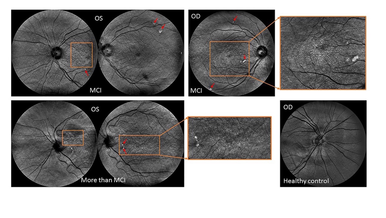
Figure 1. Retinal topographical features observed in individuals with mild cognitive impairment. Top row: Central and nasal infrared light-images obtained from a female subject (79 years old) with MCI showing extramacular features such as drusen-like regions depicted by irregularly shaped bright spots in the periphery of the superior quadrant as well as with pigment dispersion in both eyes. Bottom image: Left- Central and nasal infrared light-images obtained from a female subject (81 years old) with MCI showing tortuous vessels, extramacular features such as drusen-like regions along with pigment dispersion in the left eye. Right- Nasal infrared-light image obtained from a healthy control (71 years old). All images were acquired with the EasyScan Unit (i-Optics Corporation, The Netherlands). The EasyScan camera is a dual color confocal SLO: Infrared (785 nm) and pure green (532 nm). The different colors are related to a different penetration depth. The red arrows indicate the location of the drusen, and white spots observed at extramacular locations. The ROIs enclosed by the orange rectangles indicate the locations where pigment dispersion was observed. The green light-image is reflected at the retinal nerve fiber layer showing the vascular structure up to the 4th bifurcation. The infrared light-image is reaching the choroidal vessel layer
Collaborators: Carlos E. Mendoz-Santiesteban, M.D. (Bascom Palmer Eye Institute at the University of Miami Miller School of Medicine), Alberto R. Ramos, M.D., (Miller School Department of Neurology), Maja Kostic, M.D.(Bascom Palmer), William J. Feuer (Bascom Palmer), and Gábor Márk Somfai, M.D., Ph.D. (Semmelweis University)
Source: https://www.frontiersin.org/articles/10.3389/fphys.2020.570412/full
Sponsors: Finker-Frenkel Family Foundation;Alzheimer’s Association,;and National Institute on Aging (NIH/NIA-STTR (iScreen 2 Prevent LLC (DBA Multinostics))
Patent: DeBuc, D. “Diagnostic multi-modal biomarkers of cognitive impairment utilizing early vascular and neurogenic changes in the retina,” U.S. Patent No. 0079506 A1, March 17, 2022.
-
Innovation Center for Artificial Intelligence Regulation (ICAIR)
Software as a medical device (SaMD) are rapidly becoming an integral part of healthcare. This Centers of Excellence in Regulatory Science and Innovation (CERSI) project aims to develop autonomous frameworks for medical device regulation that utilizes advanced machine learning techniques to dynamically assess the safety and effectiveness of AI-based products in near real-time.
Collaborators: Yelena Yesha (University of Miami Department of Computer Science) and members of the ICAIR Consortium comprised by Emory University, MEDSTAR Health Research Institute, University of Arkansas for Medical Sciences, The Regents of the University of California, Upstream Vision, Vanderbilt University, West Virginia University Research Corporation, Institute of Neuromodulation, Children’s Research Institute, George Mason University, and Georgetown University
Sponsor: U.S. Food and Drug Administration
-
Kernel Flow (Kernel Inc.) for Eyes
This study is taking advantage of the eye-brain connectome and neuroscience-as-a-service in the cloud to investigate a broad range of diseases which can be evaluated by measuring degrees of activation in the visual cortex and throughout the brain. Increasing evidence suggests that retinal morphology and microvascular network conditions are indicators of various cerebrovascular, neurodegenerative, psychiatric, and developmental diseases. Visual function is often affected in cerebrovascular and neurodegenerative diseases, including ocular motility abnormalities, visual loss, contrast sensitivity changes, and defects in color vision.
As a noninvasive optical imaging technique, the Kernel Flow is a functional near-infrared spectroscopy (fNIRS) device that can be used to measure changes in hemoglobin species inside the brain. Like functional magnetic resonance imaging (fMRI), fNIRS detects these changes by measuring the absorption of light by chromophores, such as oxyhemoglobin and deoxyhemoglobin. However, fNIRS has several advantages over fMRI, including its portability and potential for long-term monitoring. The fNIRS brain computer interface uses near-infrared light, which can penetrate biological tissues more easily than visible light. This allows fNIRS to be used to measure brain activity in awake, mobile subjects. Additionally, fNIRS is relatively inexpensive and portable, making it a more accessible option for research and clinical applications. The main advantage of fNIRS over fMRI is its portability. fNIRS devices are small and lightweight, making them easy to transport and use in a variety of settings. This makes fNIRS a valuable tool for research and clinical applications where fMRI is not feasible, such as in the field or in home settings. Another advantage of fNIRS is its potential for long-term monitoring. fNIRS signals can be continuously recorded over long periods of time, making it possible to track changes in brain activity over time. This makes fNIRS a valuable tool for studying the effects of interventions on brain activity, such as drug treatments or cognitive training.
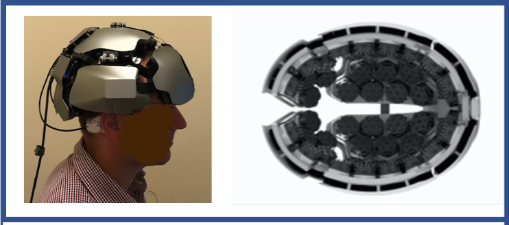
Figure 1. Kernel Flow (Kernel Inc.). The Flow 1 prototype model is a more portable/scalable device and significantly cheaper than fMRI. The device has 52 sources and 312 detectors fully integrated into a compact 2.2 Kg helmet (high channel density- whole cortex- source-detector pairs). It uses 690 nm and 850 nm laser wavelengths, and 200 Hz sampling frequency. Six active dry electrodes are also integrated into the helmet and placed close to OZ, PZ, CZ, FZ, F3, and F4 on the 10-10 grid. Sampling rate up to 1kHz per channel. The system has been designed as a small modular device that can provide real-time monitoring of tissue oxygenation in the brain as subjects take tests, perform cognitive tasks, and/or receive stimulation.
We are using the Kernel Flow system to study patients with several widespread and potentially blinding and debilitating conditions:
- Studying patients with spontaneous and evoked ocular pain to understand ocular pain pathways and develop more precise therapies.
- Studying patients with neurodegenerative diseases (e.g., AD, PD and MS) to investigate the eye-brain connections and how vision impairment affects the brain, such as in clinical cases of cognitive impairment with noncognitive neurologic findings (e.g., saccadic eye movements). This study would also reveal how brain changes might influence the outcome of potential therapeutic approaches targeting vision restoration/rehabilitation.
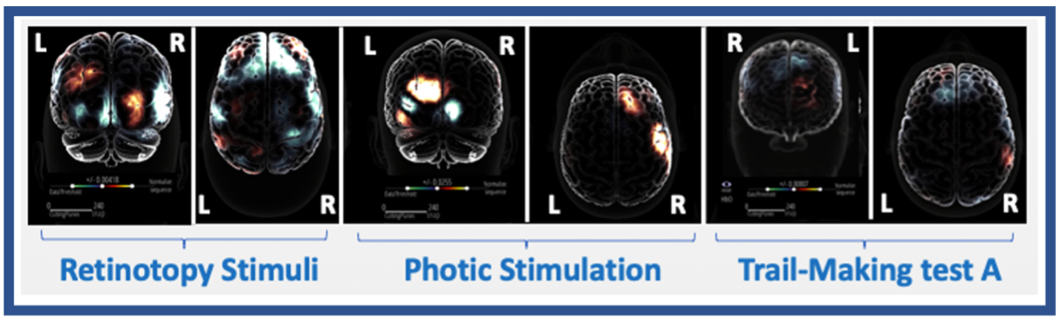
Figure 1. The fNIRS hemodynamic response (oxygenated hemoglobin - HbO) in multiple settings. Retinotopy Stimuli: Black and white wedge checkerboard stimuli were randomly presented to the four visual field (VF) quadrants of a healthy adult (28 years old, right-handed, and right- eye dominant). Activation patterns show the capability of the fNIRS technique for functional mapping of the human brain and confirm the functional asymmetry of the visual cortex previously reported by neuroimaging studies. Photic Stimulation: Cortical activation patterns during photic stimulation. The HbO increases are observed on the contralateral occipital surface during the stimulation (healthy subject, 55 years old, right-handed, and right- eye dominant, right eye stimulated with a flicker stimulus). Trail-Making Test A: Cortical activation patterns while performing the Trial-Making Test A (computer-based), a widely used neuropsychological instrument measuring executive function. The HbO increases are primarily located in the dorsolateral prefrontal cortex of the study subject (healthy subject, 28 years old, right-handed and right-eye dominant).
Collaborators: Ranya Habash, M.D., Bascom Palmer Eye Institute at the University of Miami Miller School of Medicine, Anat Galor, M.D., (Bascom Palmer), and Alberto R. Ramos, M.D., (Miller School Department of Neurology)
Sponsors: Kernel Inc.; National Institute of Health (No. P30-EY01480); and Research to Prevent Blindness, Inc.
- Studying patients with spontaneous and evoked ocular pain to understand ocular pain pathways and develop more precise therapies.
-
Novel Application for Diagnostic Assessment and Therapeutic Intervention Using Extended Reality Technology
Extended reality (XR) has moved beyond patient distraction to bring a range of proven, accessible, and transformative diagnostic and therapeutic solutions. A growing body of evidence and the potential to reduce costs, improve outcomes, and expand access to care create the momentum needed to drive widespread clinical adoption. In this project, we are developing XR-driven multi-tasking/multi-sensory paradigms to target neuroanatomical sites compromised early in AD, leading to a more robust technique in the field with potential therapeutic efficacy.
Collaborators: Ranya Habash, M.D. (Bascom Palmer Eye Institute at the University of Miami Miller School of Medicine), Kim Grinfeder, M.S., (University of Miami School of Communication) and Bryson Rudolph, M.S. (University of Miami Institute for Data Science and Computing)
Sponsor: University of Miami Provost XR Award
-
Development of Metaverse and Digital Twin Model for Intelligent Eyecare
The metaverse is the next generation of the internet, a virtual world where people can interact with each other and with digital content in a more immersive and realistic way. This project focus on the development of a digital twin of the eye using emerging technologies to enhance understanding of its intricate anatomy, physiology, and pathology. This undertaking targets medical learners, particularly at the medical student level, providing them with a comprehensive tool for studying and grasping the complexities of the eye.
A metaverse of medical technology and artificial intelligence can help to develop, prototype, evaluate, regulate, translate, and refine AI-based medical practices, especially those that involve genetics and medical imaging-guided diagnosis and therapy. Therefore, we will be applying AI/ML to create personalized digital twins of patient eyes to impact patient care and clinical outcomes.
Collaborators: Ranya Habash, M.D. (Bascom Palmer Eye Institute at the University of Miami Miller School of Medicine), Yelena Yesha, Ph.D. (University of Miami Institute for Data Science and Computing), Giselle Ricur,M.D., M.Sc., (Bascom Palmer), Kim Grinfeder (University of Miami School of Communication), M.S., and Bryson Rudolph, M.S. (University of Miami Institute for Data Science and Computing)